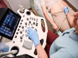
Infertility affects 1 in 6 people globally, which is more than 17 per cent of the adult population worldwide, according to a new WHO report. It showed high prevalence in all regions, high-, middle- and low-income countries, indicating that it is a major health challenge globally. Having difficulty conceiving? Don’t lose hope yet. Infertility can be treated with medicines, surgical procedures or assisted reproductive technology (ART), depending on the cause, and help you conceive.
A number of factors can cause
infertility
. Radiologists can help identify and even treat some of these underlying issues.
To understand the role of radiology in diagnosis and treatment of infertility, we connected with Dr Sunita Dube, Radiologist, Founder, Chairperson, MedscapeIndia, Aryan hospital. Excerpts follow:
Infertility is seen in both men and women owing to various causes such as abnormal sperm production or function due to undescended testicles for men,
uterine fibroids
for women, and consequences of previous infections, such as chlamydia, for both. However, some conditions in particular are the causes of infertility, including varicoceles in men and blocked fallopian tubes in women. Although, the initial assessment for an infertile couple is a physical examination that is commonly done. But radiology can help assess one’s fallopian tubes, mainly with a fluoroscopic examination.
As estimated by the Indian Society of Assisted Reproduction, more than 27 million couples in urban India are affected by infertility. AIIMS estimates that about 10-15 per cent of Indian couples are facing difficulty conceiving.
Infertility means the inability to get pregnant. In men, common fertility health issues are testicular infection, cancer, infected prostate, sperm disorders, injury to the scrotum or testicles, undescended testicles, ejaculation disorders, hormonal imbalance, and even cystic fibrosis. Fibroids, polyps, endometriosis, problems with the cervix, genetic abnormalities, irregular menstruation, and miscarriage are some of the issues in women. Age, diabetes, alcohol, smoking, stress, eating disorders, substance abuse, sexually transmitted infections (STIs), and obesity can lead to infertility. Thus, it is the need of the hour to diagnose infertility and seek timely intervention.
Imaging plays a key role in the diagnostic evaluation of female infertility. Infertility has many causes tubal and peritubal disorders, uterine disorders including mullerian duct anomalies, ovarian disorders, and cervical disorders. There is an increase in the incidence of female infertility in association with that there is an increase in demand for female imaging modalities.
Although hysterosalpingography (HSG) is the gold standard investigation, ultrasound sonography (USG) is the first line of investigation for female infertility, easily carried out with painless non radiation.
Sonosalpingography (SSG) or Sion test distinguishes uterine synechiae, endometrial polyps, and submucosal leiomyomas. Pelvic MRI can help detect
endometriosis
, adenomyosis, leiomyomas, and Mullerian duct anomalies. Cervical stenosis is identified in HSG as difficulty or failure in cervical cannulation. Ovarian disorders are usually diagnosed by USG.
A multimodality imaging approach is necessary to identify the exact cause of infertility, following which appropriate management can be initiated.
In women, radiology can help assess one’s fallopian tubes, mainly with a fluoroscopic examination. Also, one can get the contrast ultrasound done so that there is no radiation and can assess the uterus at the same time. However, most people go for fluoroscopic technique because it provides more detail and allows you to potentially undertake recanalization.
A hysterosalpingography means an X-ray to check the woman’s uterus (womb) and fallopian tubes (structures that transport eggs from the ovaries to the uterus) of the woman by injecting a radio-opaque dye in the uterine cavity. The type of X-ray used is called fluoroscopy which allows real time visualization of dye passing through the uterus and thus detects blockage in your fallopian tubes or other structural abnormalities in your uterus. Hysterosalpingography may be also referred to as uterosalpingography. Apart from that, this test can help to detect scarred tissue in the uterus, polyps, uterine tumors, and uterine fibroids


 Driving Naari Programme launched in Chandigarh


